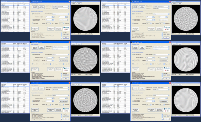

With the Dr. Probe Light software one can calculate and display electron probes, electron wave functions, phase plates, and ronchigram images of amorphous objects. A large number of parameters allow you to specify conditions close to realistic situations with scanning transmission electron microscopes (STEM). All parameters are setup within a graphical user interface. By fast calculation and display of the various probe functions, the effects of parameter changes are made visible in real-time.
Essentially, Dr. Probe Light should be seen as a learning tool for people working with aberration correction at scanning transmission electron microscopes. You can learn how the different microscope parameters affect the electron probe geometry at the object plane. You may also learn the effects of the optical aberrations in ronchigram images of amorphous objects, which are frequently used as a visual help for manual aberration correction of a STEM.
As a special feature Dr. Probe Light provides a computer-controlled ronchigram aberration correction training (RACT) on the basis of ronchigram images of a thin amorphous object. The main purpose of this feature is to help you in learning how coherent aberrations like twofold astigmatism and coma govern ronchigram images and how you can eliminate these aberrations as fast as possible. Your training success is evaluated by a training score, which reflects your skill in quickly adjusting aberration corrected states of a STEM.
Dr. Probe Light is a sub-unit of the program package Dr. Probe comprising functions related to the probe forming in STEM. In addition, Dr. Probe contains a complete multislice simulation for the evaluation of the electron wave propagation through an object and for the calculation of STEM images. Dr. Probe is developed at the Forschungszentrum Jülich GmbH (Jülich, Germany) since 2007. If you want to learn more, please feel free to contact the developer of the program, Juri Barthel.
