Sample Visualization


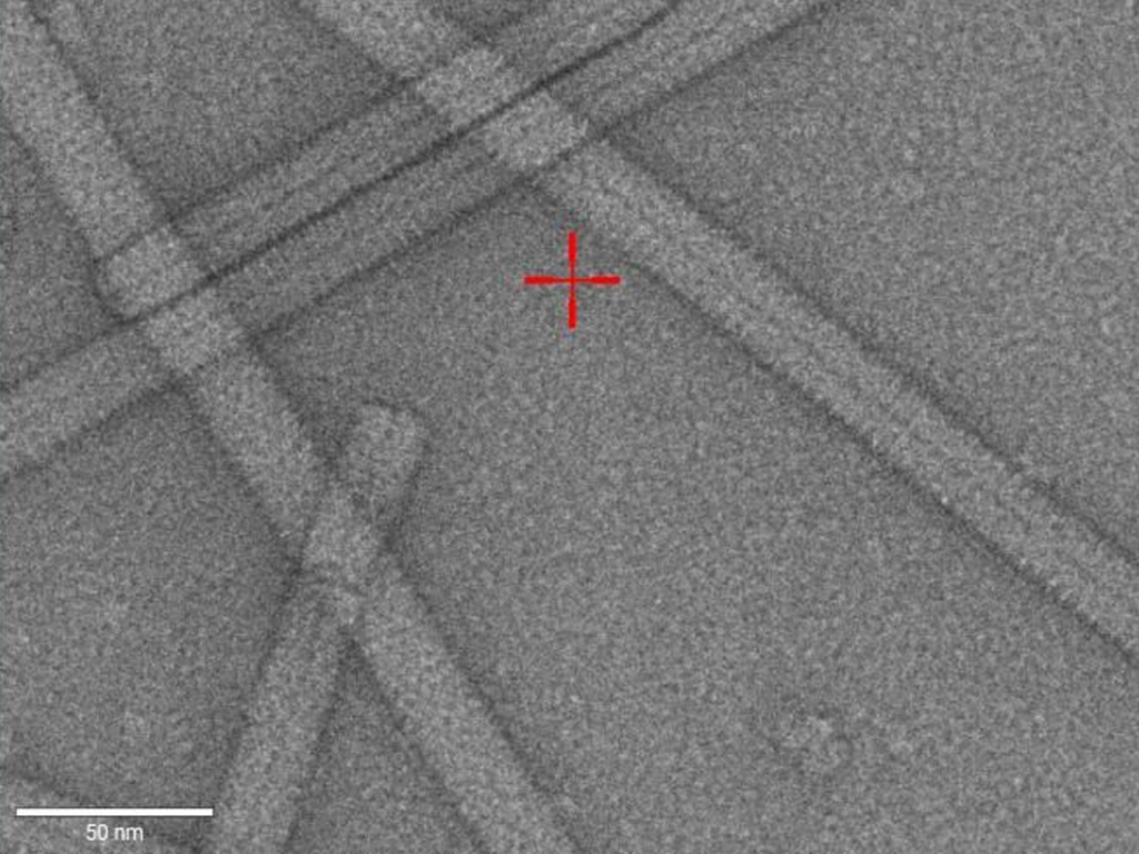
The aim of the initial visualization is to check purity and homogeneity of the sample and it further provides information about sample morphology and dimensions.


Negatively stained samples

Purified proteins, filaments, viruses and other large assemblies can be rapidly visualized and characterized by negative stain electron microscopy using e.g. our Talos 120 microscope. Samples are often initially imaged using negative staining EM for optimizing cryogenic single-particle and tomography specimens

Micrograph of tobacco mosaic virus (TMV) particles as viewed at 92 kX nominal magnification in our room temperature Talos 120 kV.

Vitrified cryo-samples

We offer the visualization of vitrified cryo-samples, e.g. protein complex and lipid mixtures for sample characterization. This visualization can also be part of the screening step to assess the sample feasibility for the single-particle data acquisition on cryo-EM. The plunge-frozen sample can be shipped to the ER-C or be vitrified on site using one of our sample preparation instruments like VitrobotTM, VitroJetTM, Manual or LeicaTM Plunge freezer. A small data set can be collected for initial sample characterization that may shed light on the suitability for high-resolution analysis.

Micrograph of vitrified tobacco mosaic virus (TMV) particles as viewed at 100 kX nominal magnification in our Talos Arctica 200 kV operated at cryo-conditions.

Samples often need multiple rounds of initial screening until requirements for high-resolution cryo-EM are met. These optimization includes adjustments of both, sample conditions (pH, buffer, additives) and blotting conditions (grid type, blotting time, concentration, vitrification procedure).