Workshop Details
For sustainability reasons, this information is available only as digital version
CORRIDOR: Life Sciences
Mentors
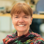
Nicole Amberg

________________________________________________
I have a long-lasting passion for patient-relevant research. I am particularly curios on how stem cells interact with their cellular environment and how this interaction shapes stem cell identity and fate in health and disease. During my PhD in the lab of Maria Sibilia at the Medical University Vienna, I was investigating the complex interaction between tumors, the immune system and stem cells in the skin. Due to my interest in cancer-microenvironment interactions, I have contributed to several influential papers of the Sibilia lab, assessing tumor-immune cell interactions in the liver and intestine. During all of these studies, I realized that one of the great limitations of cancer research is to mimic a patient-relevant somatic cell mosaicism by inducing and investigating a targeted gene mutation in single stem cells within a healthy tissue. One of the very few genetic models allowing such manipulation is Mosaic Analysis with Double Markers (MADM). In order to gain expertise in the application of MADM I thus decided to join the lab of Simon Hippenmeyer at the Institute of Science and Technology Austria for my Postdoc. At the same time, I took the chance to broaden my scientific knowledge by changing fields to developmental neuroscience. In my postdoctoral FWF-funded project, I set out to investigate the function of the epigenetic repressor Polycomb Repressive Complex (PRC)2 in neocortical development. Due to its cell-intrinsic mode of action, PRC2 activity has generally been thought to be a cell-autonomous one. Yet, stem cells are embedded within a complex cellular environment and it was unknown, whether global tissue-wide mechanisms shape PRC2 function in individual progenitors. To this end, I have contrasted a single-cell mutant with a whole-tissue ablation paradigm and genetically dissected the interplay of PRC2 cell-autonomous and global tissue requirement. I discovered that PRC2 is not cell-autonomously required for neural progenitor state determination and neuron production. Overall, my work made the novel discovery that PRC2-dependent transcriptional control regulating progenitor cell lineage progression strongly depends on the genetic tissue-wide landscape and cellular environment. In September 2022, I joined the Department of Neurology at the Medical University Vienna as a young PI. Being positioned in a clinical setting grants me direct access to precious patient material in our neurobiobank and provides me with excellent state-of-the-art expertise of the medical staff. My lab is interested how a brain of correct size and cell composition forms during development and how pediatric brain tumors arise. In particularly, we are focusing on:
(1) PRC2 function in human brain development.
(2) patient-derived iPSCs and cerebral organoids to understand the cell-autonomous and non-cell-autonomous mechanisms of pediatric brain tumor development.
(3) Detailed characterizations of human brain malformations.
In addition, I am a strong advocate for women in science and a known science communicator in Austria. I give public speeches, have TV appearances in a popular TV show and have also successfully gained two FWF grants for science communication projects. The latest will develop a narrative audiostory and an NFC-tagged brain figurine and target 8-10 year old kids.
Bridget Carragher

________________________________________________
Bridget Carragher received her Ph.D. in Biophysics from the University of Chicago in 1987. She worked in a variety of positions, both in industry and academia before moving to the New York Structural Biology Center in 2015 to lead the Simons Electron Microscopy Center (SEMC), together with Clint Potter. While at SEMC, Bridget and Clint directed the National Resource for Automated Molecular Microscopy (NRAMM), the National Center for CryoEM Access and Training (NCCAT), the National Center for In-situ Tomographic Ultramicroscopy (NCITU), and the Simons Machine Learning Center (SMLC). They also founded the company NanoImaging Services in2007. Bridget moved to her current role as Founding Technical Director of the Chan Zuckerberg Imaging Institute (CZII) in January 2023. The mission of CZII is to enable deep insights into the architecture of complex biological systems, at the molecular level, through the development and application of novel imaging technologies. The initial grand challenge is to develop technologies and methodologies to image the molecular architecture of the cell to near atomic resolution using cryo electron tomography.
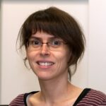
Marta Carroni

________________________________________________
I am Marta Carroni, I come from Italy, more specifically from a little town in the mountainous part of the island of Sardinia. The Sardinian society I grew up in is a strong matriarchal one and I get a strong feeling of freedom when I think of my grandparents from both sides. Living on an island can be nevertheless quite limiting and I left Sardinia at 19 years old to take the university studies in Pisa, in the mainland. I moved to Madrid for one year Erasmus followed by the master studies. I didn’t come back to Italy ever since. I am a convinced European and I think that common means of communication, like science or shared languages, unite people. I moved to London for my PhD and postdoc and there in the UK, where I lived for 10 years, I had great female mentors: my PhD supervisor Silvia Onesti and my postdoc supervisor Helen Saibil. I have been also surrounded by male mentors with open and advanced views about equity, such as Peter Brick and Steve Curry. After London, around 9 years ago, I moved to Stockholm in Sweden to, eventually become the head of the national cryo-EM infrastructure. Here I got a very strong support from a male mentor, Gunnar von Hejine. I never really felt I had to face major gender discriminatory situations. However, as I became older and maybe more mature, I realized that there are deeply embedded cultural bias that make it harder for a woman to affirm herself in the academic environment. Being self-confident is highly prized in the academic environment, even in the scientific one. Somehow, males are still educated differently from females, to be bolder and less caring of other people’s opinion. This is of coincidence that eating disorders are more spread among teenage girls than boys. This is of course a rough generalization, but it is also quite common to assume that, if a man and a woman are in the same age and similar positions, the boss is the man. Somehow, to fight these culturally-rooted unconscious bias, it is important to actively bring up discussions whenever unfair situations are detected. I actually noticed that there is another type of discrimination that goes under the radar. I would call it the geographical discrimination, by which people coming from rich countries almost automatically receive better recognition than those coming from poorer countries. Science should become more blind in many aspects.
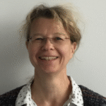
Katharina Hipp

________________________________________________
Katharina Hipp is Head of the Electron Microscopy Facility at the Max Planck Institute for Biology Tübingen, Germany, where research covers a wide range of biological samples from molecular, complex organism down to single cells and purified macromolecular complexes. Originally trained in molecular biology and plant virology, she became fascinated by electron microscopy early on already during her studies. As a PostDoc she joined Bettna Böttcher’s group in the Structural and Computational Biology Unit at EMBL Heidelberg, Germany, diving into single particle cryoEM. She continued with cryo-EM at the University of Edinburgh, School of Biological Sciences, UK, as a PostDoc and SULSA cryoEM technologist. After returning to Germany, she started as a group leader in Holger Jeske’s lab at the University of Stuttgart studying plant viruses with a focus on structural biology and cellular biology. She completed this work with her habilitation in Molecular Biology at the University of Stuttgart, Germany, in 2020. Currently, she is the President of the German Society for Electron Microscopy that brings together physicists, material scientists and scientists from the life sciences. She is married and has two children.
Catherine Venien-Bryan

________________________________________________
Catherine Vénien-Bryan obtained her PhD in Molecular Biophysics at the University Pierre et Marie Curie , Paris (Sorbonne Université). She discovered cryogenic-electron microscopy (cryo-EM) applied to structural Biology during her first post-doc at EMBL Heidelberg Germany (Structural Biology program). This was followed by an ECC Bridge fellowship at the University of Oxford, Biochemistry Department, which enabled her to deepen her knowledge in the field of membrane proteins. In 1993, she was recruited as Assistant Professor at the University of Grenoble, where she focused on the development of biophysical techniques for the study of proteins. She then returned to Oxford University (2000-2012) to take up a University Lecturer position in the Biochemistry Department where she was involved in the Structure and Dynamics of signaling proteins. In 2012, she accepted the position of Professor of Biophysics at Sorbonne Université and Group Leader at IMPMC. Her research focuses on the structure and function of human potassium channels. Many signals in the cell are conveyed by interacting protein molecules. How do protein-protein interactions lead to a response? The most likely explanation is through changes in structure. With her group; she studies protein-protein interactions in the control of signaling processes using cryo-EM and combining the results with information from X-ray diffraction and biophysical studies.
Peijun Zhang

________________________________________________
Peijun Zhang is a Professor of Structural Biology in the Nuffield Department of Medicine at the University of Oxford and the founding director of eBIC (the UK National Electron Bio-imaging Centre) at the Diamond Light Source. She obtained B.S. in Electrical Engineering and M.S. in Solid State Physics from Nanjing University, and Ph.D. in Biophysics and Physiology from University Virginia. She was a post-doctoral fellow and subsequently a staff scientist at the National Cancer Institute, NIH, and then an Assistant Professor and tenured Associate Professor at the University of Pittsburgh School of Medicine. She joined Oxford and Diamond Light Source in 2016. Professor Zhang is a leading expert in the fields cryoEM and cryo-electron tomography (cryoET) of macromolecular complexes and assemblies, especially investigating these in situ in the native cellular context. Her research is aimed at an integrated and atomistic understanding of molecular mechanisms of viral and bacterial infections by developing and applying novel technologies for high-resolution cryoEM and cryoET. Her current research efforts focus on HIV-1 and SARS-CoV-2 infections using multi-scale correlative structural biology and bacterial chemotaxis signalling pathways using time-resolved cryoEM and cryoET. She has been awarded with prestigious grants such as Wellcome Investigators Award, ERC AdG grant, and Wellcome Discovery Award. She is an elected member of the European Molecular Biology Organization (EMBO) and has received many Honors and Awards, including The Senior Vice Chancellor’s Award, Carnegie Science Emerging Female Scientist Award and Distinguished Research Career Award.
Industry
Thermo Fisher Scientific – Life Sciences

________________________________________________
Dr. Lingbo Yu has been working in electron microscopy for 20 years. She started as a Ph.D. student under the supervision of Prof. Michael Radermacher. Her research focuses on alignment and classification algorithms for subtomogram averaging. After graduation, she joined FEI-NIH living lab project, and worked on automated data collection. Upon finishing of the project, she took the product management role for various products, including Falcon direct electron detector, micro electron diffraction, Krios and Tundra cryo-TEMs. She is now a Sr. product marketing manager, mainly responsible for Glacios 200kV cryo-TEM.
Thermo Fisher Scientific are proud of our Mission: To enable our customers to make the world healthier, cleaner and safer. Through our electron microscopy solutions and expertise, we help customers accelerate innovation and enhance productivity across the life sciences. We offer a broad range of products covering all imaging scales from near-atomic resolution molecular data to tissue and whole cell ultrastructure. By revealing molecular detail at biologically relevant resolutions, we enable scientific discoveries and breakthrough drug discovery. At Thermo Fisher Scientific, Diversity & Inclusion is vital to the future success of our mission. Our Women’s Employee Resource Group specifically is committed to making Thermo Fisher Scientific one of the world’s most admired companies by fostering the advancement of women and building a corporate culture in which women employees are recruited, valued, developed, retained, and promoted globally.
CryoCloud


________________________________________________
Dr. Ieva Drulyte is a structural biologist with over a decade of experience in cryo-EM, spanning both academic and industry settings. She holds a PhD in structural enzymology from the University of Leeds and has worked at the interface of science and technology throughout her career. While at Thermo Fisher Scientific, she collaborated with leading pharmaceutical and biotech companies on structure-based drug discovery projects across a range of modalities – including small molecules, peptides, degraders, DARPins, and antibodies – contributing to 17 publications, 25 PDB entries, and 47 EMDB depositions. In her current role as Commercial Director at CryoCloud, Ieva leads sales, customer success, and strategic collaborations. She works closely with structural biologists to help them make the most of the CryoCloud platform for cryo-EM data processing and management. Passionate about bridging scientific innovation with real-world application, Ieva is committed to supporting researchers and advancing structural biology through more efficient, data-driven workflows.
Robert Englmeier, PhD is the Co‑Founder & CEO of CryoCloud, a cloud-based platform for cryo‑EM data analysis. Originally from Munich, he moved to the Netherlands in 2016 to pursue a PhD in cryo‑electron microscopy at Utrecht University, where he focused on mitochondrial protein synthesis using cryo-ET. In mid‑2021, while finishing his doctoral work, Robert joined the BCF BioBusiness Summer School. Inspired by the entrepreneurial talks, he pitched the concept of CryoCloud and entered the UtrechtInc Incubator program where he co-founded CryoCloud with two other co-founders. After graduating in June 2022, they launched a working cloud application and accumulated hundreds of active analysis hours by early 2023. Under his leadership, CryoCloud has raised over €2.8 million in funding, enabling the development of proprietary ML-based and classical algorithms, cryo-ET workflows, and highest security standards including ISO‑27001 compliance. The platform now supports users in more than 25 countries and serves academia, biotech and pharma clients. A structural biologist and technologist at heart with over ten years of cryo‑EM experience, Robert is committed to enabling and democratizing automated, high-throughput cryo‑EM via the cloud and the development of cutting-edge algorithms.
CryoCloud develops cloud-native software solutions for cryo-EM data analysis, storage and management. CryoCloud’s web-app enables scientists to analyze data on dozens of GPUs in minutes, while automation and collaborative features as well as proprietary algorithms improve the efficiency and quality of results. The CryoCloud web-app eliminates the need for hardware setup and maintenance, and provides the highest information security standards verified by external parties. CryoCloud’s mission is to automate cryo-EM data analysis and provide scientists with the simplest and fastest way to determine protein structures at the highest quality.
Bruker

________________________________________________
Meiken Falke, Global Product Manager at the Bruker Nano GmbH Berlin, is engaged in improving instrumentation for structure analysis on the nanoscale in materials and life science. She has over 30 years of experience in electron microscopy. Meiken joined Bruker in 2008 to focus on the development and application of detector systems for energy dispersive X-ray spectroscopy and the respective business development. Before that she worked as lecturer researching how materials macroscopic behavior depends on their nanoscale properties. Between 2002 and 2005, Meiken was one of the first postdocs at the SuperSTEM Laboratory in Daresbury UK supporting the application of aberration correction in transmission electron microscopy and the setup of this international user facility.
The Bruker Nano GmbH, part of Bruker Corporation, develops, manufactures, and markets systems for elemental and structural analysis on the micro and nano scale. Our unique range of analytical tools include EDS, WDS, EBSD and micro-XRF instruments for the electron microscope and offer the most comprehensive compositional and structural analysis of materials available today. The full integration of all these techniques into the ESPRIT software allows you to easily combine data obtained by these complementary methods for best results. On top we offer a variety of benchtop micro-X-ray fluorescence (micro-XRF) spectrometers for spatially resolved composition analysis and total reflection X-ray fluorescence (TXRF) instruments for trace element analysis for a multitude of applications. Our handheld XRF analyzers as well as the mobile/portable countertop XRF analyzer, enabling non-destructive and on-site element analysis complete the portfolio. The Bruker Nano GmbH portfolio covers a variety of applications such as material science, life science, geo science, other as well as many industrial applications.
